When people talk about “gastritis” in everyday conversation, they often use the term to describe any stomach discomfort. However, gastritis is the correct medical term only when inflammation of the stomach lining has been confirmed microscopically. Today, gastritis is classified into several types: A, B, C, and D.
In this article, we focus on type B gastritis, the most common form, which is caused by the bacterium Helicobacter pylori. We will also explain why treating this infection is so important.
Briefly, type A gastritis is an autoimmune condition that occurs relatively rarely, mainly in older adults. Type C gastritis is caused by irritants such as alcohol and smoking, which damage the stomach lining. Type D gastritis encompasses rare, recently described diseases of the stomach lining, including collagenous gastritis, eosinophilic gastritis, vasculitic gastritis, and other uncommon conditions.
What is Helicobacter pylori and why is it important?
H. pylori is a bacterium that can cause an infection with or without noticeable symptoms. Regardless of whether symptoms are present, the infection causes chronic active inflammation of the stomach lining. Over time, this inflammation may lead to serious complications.

The most important complications include:
- Gastric or duodenal ulcers (which may bleed)
- Gastric cancer (stomach cancer)
- MALT lymphoma (a rare lymphoma of the stomach lining)
It is well established that H. pylori infection increases the risk of stomach cancer by two to three times compared with people who are not infected. Although the infection remains common worldwide, its prevalence has gradually declined over recent decades, particularly in developed countries.
How is H. pylori transmitted?
The exact route of transmission has not yet been fully clarified. It is believed that the bacterium may spread via saliva, gastric contents, the fecal-oral route, or a combination of these mechanisms. Transmission through contaminated food or water has also been considered, although this is uncommon in developed countries.
It is also important to note that genetic susceptibility plays a role—some individuals appear to acquire the infection more easily than others.
Can the infection disappear on its own?
Yes, but this is uncommon. Spontaneous clearance may occasionally occur in infants and very young children. However, spontaneous resolution is extremely rare in preschool-aged children and adults. In clinical practice, once the infection is established, it usually persists until eradication therapy is administered.
How is the infection diagnosed?
Both invasive and non-invasive diagnostic methods are available.
Invasive methods (performed during gastroscopy)
- Histological examination of gastric biopsy samples
- Rapid urease test
- Bacterial culture
- PCR analysis
Non-invasive methods
- ¹³C urea breath test
- Stool antigen test for H. pylori
The most reliable diagnosis is achieved by combining two different diagnostic methods, although this is not always necessary in routine clinical practice. For example, if a duodenal ulcer is identified during gastroscopy and one diagnostic test is positive, this is generally considered sufficient evidence of H. pylori infection.
Which diagnostic method is the best?
There is no single diagnostic method that is best for every patient. The choice depends on the clinical situation and whether gastroscopy is being performed. Invasive methods—including histology, the rapid urease test, bacterial culture, and PCR analysis—require gastric biopsy samples obtained during gastroscopy. Among the non-invasive methods, the ¹³C urea breath test and the stool antigen test are the most commonly used. The ¹³C urea breath test is considered one of the most accurate non-invasive tests for detecting active H. pylori infection.

What can cause false-negative test results?
Several factors may temporarily suppress the bacterium and lead to false-negative results:
- Proton pump inhibitor (PPI) therapy (such as pantoprazole or esomeprazole)
- Recent antibiotic treatment
- Upper gastrointestinal bleeding
- Previous stomach surgery
- Advanced atrophic gastritis or
- intestinal metaplasia Gastric cancer or MALT lymphoma For these reasons, repeat testing may sometimes be necessary.
What does the examination involve?
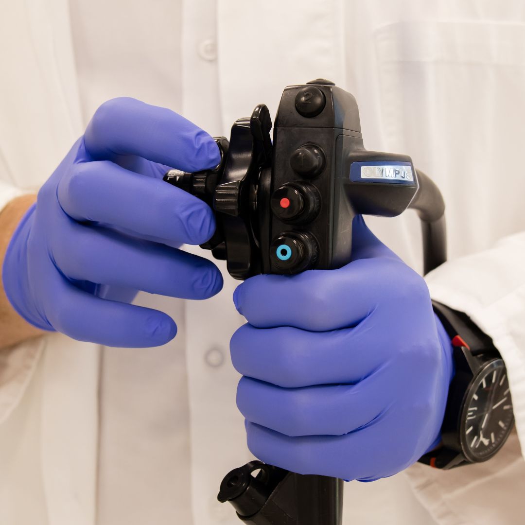
During gastroscopy, biopsy samples are routinely taken from the stomach lining to test for H. pylori. Because this bacterium causes type B gastritis and increases the risk of stomach cancer, treatment is recommended whenever the infection is confirmed.
If a gastric or duodenal ulcer is also detected, a follow-up gastroscopy is often recommended after treatment to confirm ulcer healing and determine whether additional biopsy samples are needed to exclude malignant disease.
You can book your initial gastroenterology consultation at IMC Priora, where Dr. Matej Plažanin and the Department of Gastroenterology will evaluate you.

