Mona Lisa Dx Biobot
Mona Lisa Dx Biobot
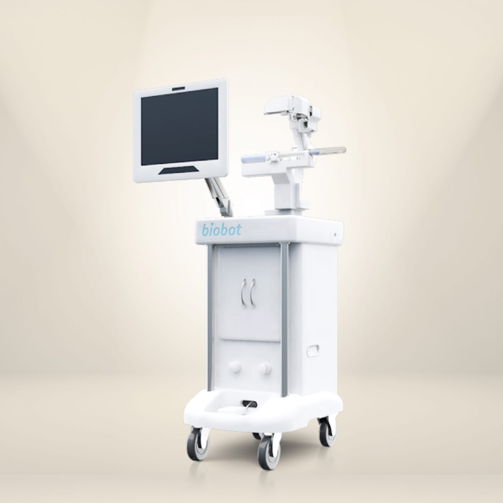
Mona Lisa Dx Biobot is revolutionizing prostate biopsy. This cutting-edge robotic technology offers a more precise, minimally invasive, and streamlined approach to prostate cancer detection, improving accuracy and minimizing risks for both doctors and patients.
By enabling a potentially safer and more accurate diagnosis of prostate cancer, Mona Lisa represents a significant advancement in prostate biopsy technology and could revolutionize early detection and management of the disease.
Minimally invasive procedure with reduced discomfort
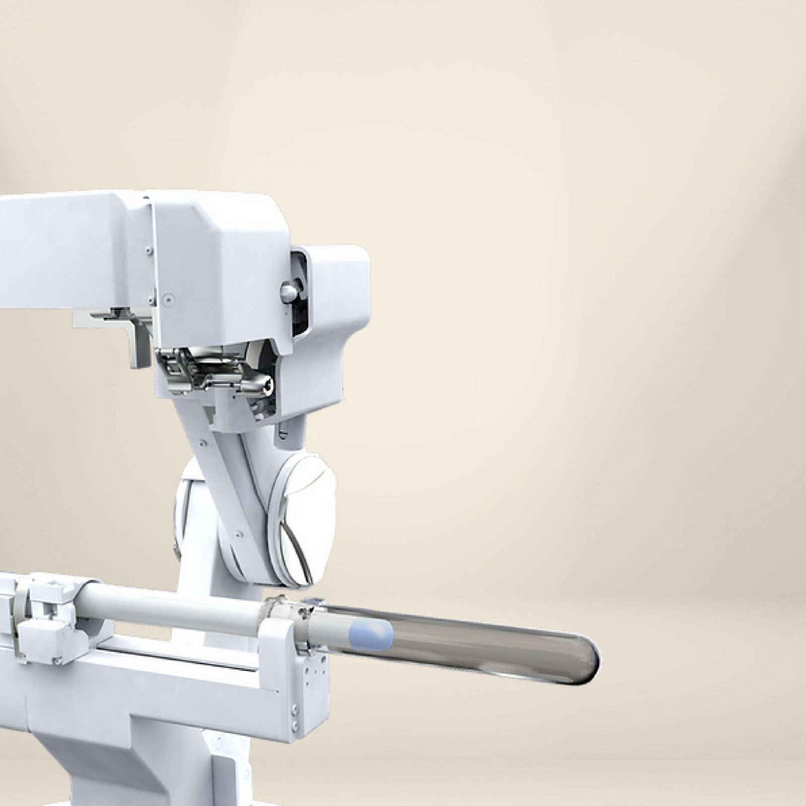
The Mona Lisa robot redefines prostate biopsy, prioritizing both patient comfort and surgical control. Mona Lisa’s robotic system empowers urologists by providing enhanced stability and precision, greatly lowering the possibility of human error. This translates to a more targeted approach, minimizing unnecessary sampling and reducing procedure time. Mona Lisa offers a revolutionary and safer experience for prostate biopsy patients.

Fusion biopsy enhances precision for improved cancer detection
Traditional biopsy methods often miss cancerous areas due to limited ultrasound imaging and random sample collection. The cutting-edge Mona Lisa robot utilizes fusion biopsy. The procedure includes a surgeon analyzing pre-biopsy MRI scans overlapped with real-time ultrasound images of the prostate scanned in the operating room. Additionally, the urologist can use the fused scans to create a 3D sonographic model of the prostate section, providing another dimension for visualizing affected areas. Furthermore, the robotic arm guided by MRI data precisely positions the biopsy needle, ensuring tissue samples are collected from the targeted areas.
Mona Lisa’s fusion biopsy technology enables exceptional visualization and highly targeted core sampling, significantly improving cancer detection rates.

Benefits for patients: comfort and fast recovery
Patients undergoing a Mona Lisa biopsy experience several advantages. The minimally invasive procedure requires only two small skin punctures, reducing discomfort and leading to faster recovery times. Furthermore, unlike traditional methods, it avoids the rectum, significantly reducing infection risk. Its unique design minimizes tissue trauma and discomfort during the procedure.
Robotic guidance and probe stabilization techniques used by the Mona Lisa may further improve precision and efficacy compared to traditional methods.
Streamlined Workflow for Efficient Biopsy
-
1. Customized Biopsy Plan
The radiologist identifies suspicious areas on the pre-biopsy MRI scans and creates a customized biopsy plan.
-
2. Imaging the Prostate
The urologist guides a real-time ultrasound probe and scans the patient's prostate. Specialized software fuses the live ultrasound data with pre-biopsy MRI scans. This creates a highly detailed 3D model of the prostate. The urologist uses the model to calculate the prostate biopsy coordinates and the Mona Lisa robot adjusts the biopsy needle accordingly and collects samples from target areas.
-
3. Precision Biopsy
A perineal biopsy is a medical procedure used to obtain tissue samples from the prostate through the area between the anus and the scrotum, known as the perineum. This method provides accurate tissue samples for diagnosing prostate cancer or other prostate abnormalities. A perineal prostate biopsy using the Mona Lisa system enables exact sampling through the perineum with minimal risk of infection. The Mona Lisa robot employs advanced needle-guidance techniques, reducing the need for multiple punctures and ensuring patient comfort. The procedure is quick and safe, aiming for the timely detection of potential prostate abnormalities.
-
4. Data analysis
Following the procedure, a comprehensive report with clinical data and 3D images is automatically generated for further analysis.
Consultation
To determine if you are the right candidate for a biopsy, we invite you to contact us and schedule a consultation. During the consultation, you will have the opportunity to discuss the procedure with your doctor and get answers to all your questions.
Our team will carefully guide you and provide you with all the necessary information for proper preparation for the biopsy. We will provide you with medical support and personalized guidance for a smooth experience.

