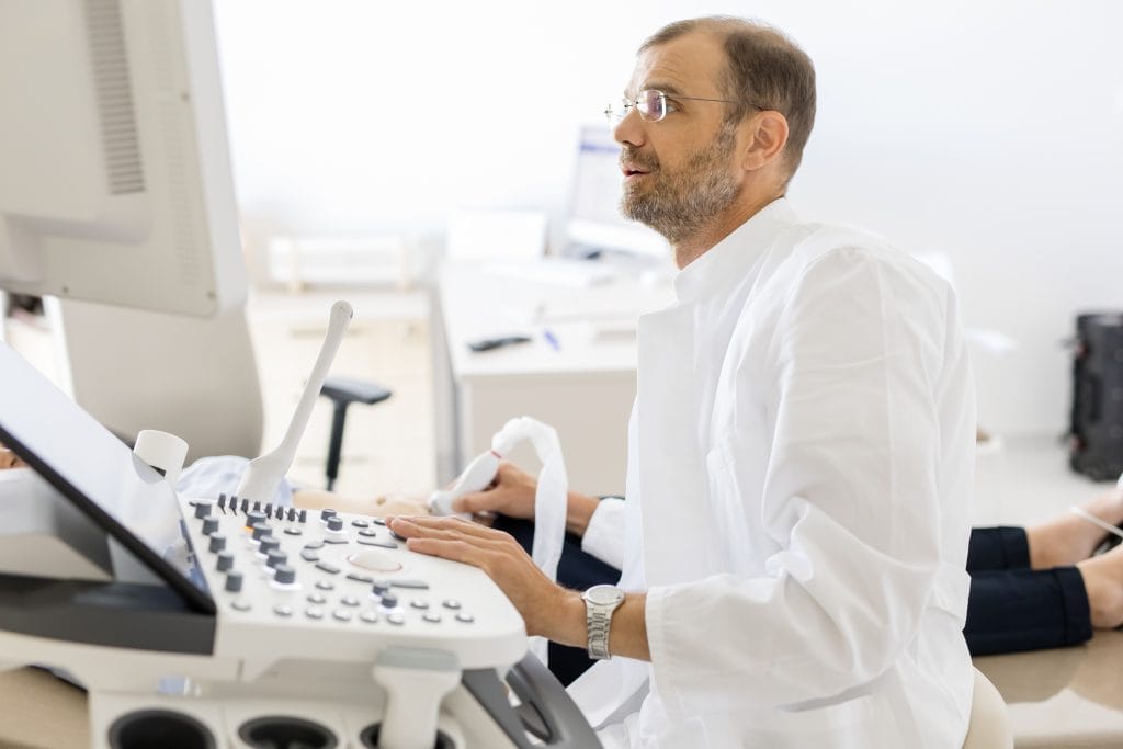
The hysteroscope, equipped with a camera and light at its tip, allows the doctor to precisely locate and remove a polyp or myoma, explains Dr. Dimitrije Milojković, Head of the Gynecology Department at IMC Priora.
Hysteroscopy is a minimally invasive procedure in which the gynecologist uses an endoscope – the hysteroscope – to insert a camera into the uterus in order to detect abnormalities that may cause, for example, heavy bleeding or infertility. These changes can then be removed during the same procedure.
The hysteroscope does not damage the uterus
“Thanks to the camera and illumination at its tip, the hysteroscope enables the physician to precisely identify and remove a polyp or myoma. It is important to emphasize that during a hysteroscopic procedure, other parts of the uterine cavity or its lining are not damaged,”
says Dr. Dimitrije Milojković, Head of the Gynecology Department at IMC Priora.
Hysteroscopy is performed when an ultrasound detects or raises suspicion of a polyp or myoma that causes irregular or heavy bleeding and subsequent anemia, or could interfere with a future pregnancy — making its timely removal highly recommended.
Patients are admitted on the day of the procedure. In the case of smaller growths, anesthesia is not always required, allowing the patient to remain awake and follow the procedure in real time. The thin hysteroscope can be inserted without dilating the cervix — which is normally the most painful part of the process — so the procedure can be performed comfortably, even without sedation. However, anesthesia remains available for those who prefer it.

Dr. Milojković: hysteroscopy is an exceptionally objective method
The uterus is gently filled with a special liquid to expand it and allow a clear view of the uterine cavity, enabling the doctor to immediately remove any detected lesions. This fluid may cause mild cramping in the lower abdomen, so the patient receives an analgesic and, if necessary, a local anesthetic around the cervix before the procedure.
“It is an exceptionally objective method — more reliable than an ultrasound examination. The angled camera improves visibility of the uterine cavity and provides a more precise assessment. From a diagnostic standpoint, hysteroscopy is also recommended in cases of hormonal imbalance, to collect a sample of the uterine lining for further analysis and treatment planning. In infertility cases, hysteroscopy is used to evaluate the uterine cavity and obtain samples for histological examination,”
explains Dr. Milojković.
IMC Priora places great emphasis on advanced medical technology to provide the highest standard of care for its patients. In the field of hysteroscopy, IMC Priora uses the most modern system available, capable of removing larger polyps and myomas without anesthesia, thanks to the device’s ability to fragment and remove tissue without the need for cervical dilation.
