What is hysteroscopy?
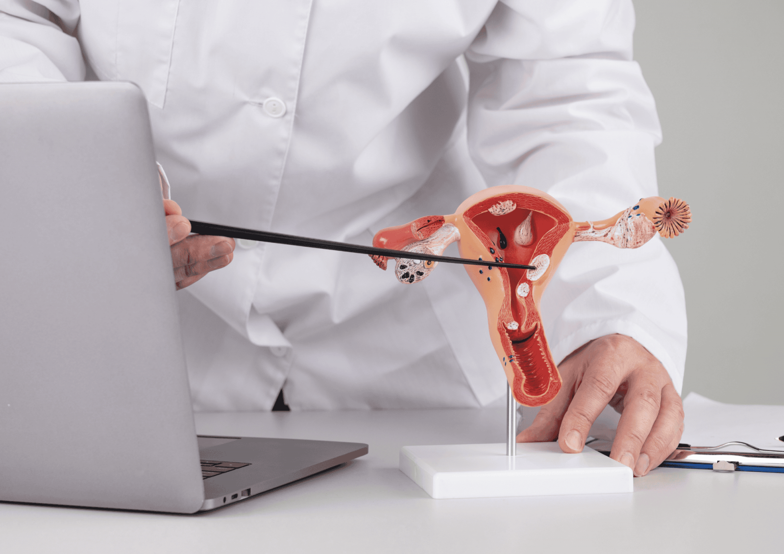
Hysteroscopy is a minimally invasive diagnostic and therapeutic procedure in which a gynecologist uses an endoscope (a hysteroscope) to examine the inside of the uterus and, if necessary, immediately remove abnormalities such as polyps or fibroids.
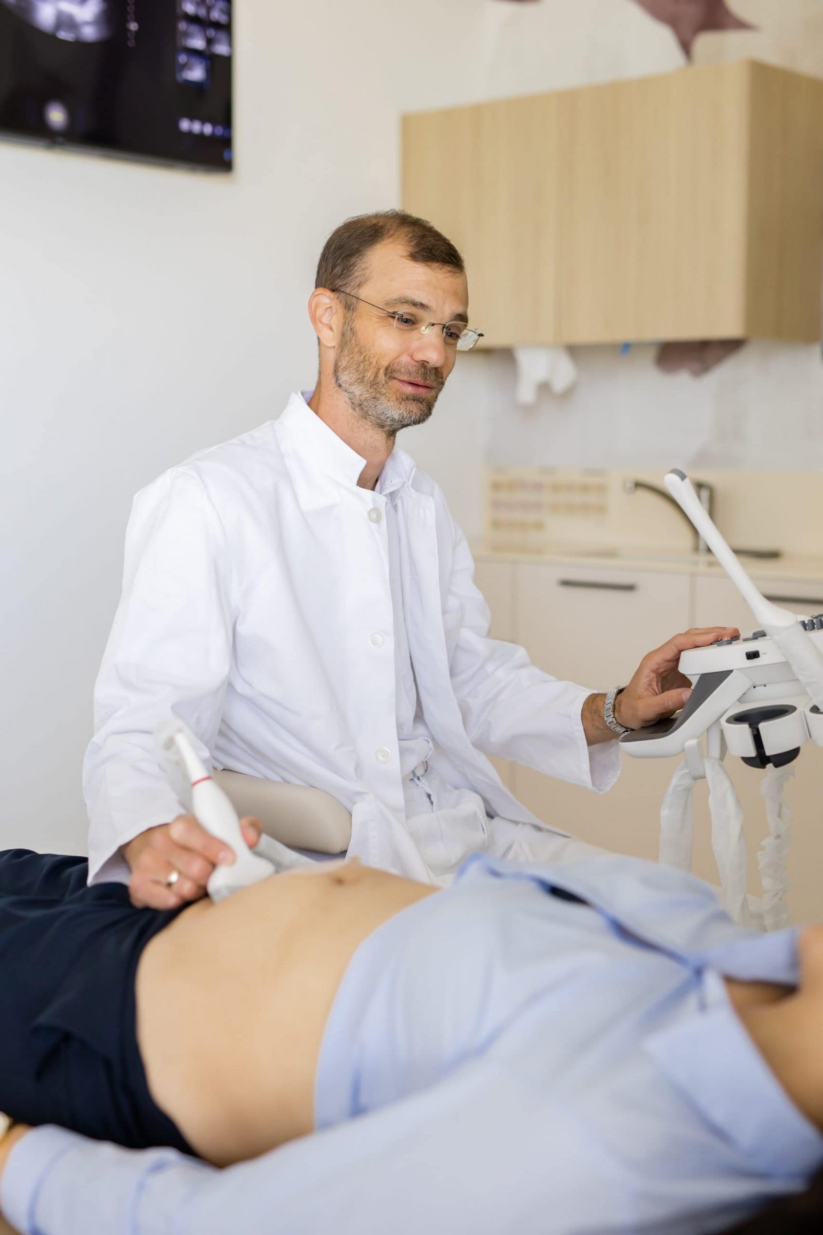
The hysteroscope is a thin instrument with a camera and light at its tip. Gynecology specialist Dr. Dimitrije Milojković performs the procedure.
When is hysteroscopy recommended?
The procedure is most often performed when:
– an ultrasound shows or suggests a polyp or fibroid,
– a woman experiences irregular or heavy bleeding,
– anemia is present due to blood loss,
– There is suspicion that a growth may interfere with pregnancy or embryo implantation,
– There is a need to take a sample of the uterine lining (biopsy).

How is hysteroscopy performed?
– a thin hysteroscope with a camera and light is inserted into the uterus,
– the uterus is filled with a special fluid to expand it and allow a better view,
– if an abnormality is detected (e.g., a polyp or fibroid), it can be removed immediately using the same instrument,
– For minor changes, anesthesia is not required, and the patient can watch the procedure.
– If needed, local or general anesthesia may be applied.

Advantages of hysteroscopy
– an exact and reliable method, more accurate than ultrasound in assessing the uterine cavity,
– quick recovery – most patients go home the same day,
– minimal tissue damage – surrounding lining remains intact,
– allows simultaneous diagnosis and treatment,
– The latest device on the market enables removal of larger polyps and fibroids without anesthesia, thanks to the ability to fragment the removed tissue. This avoids dilation of the cervix, which is otherwise painful.
The procedure may cause mild discomfort; the patient might feel slight cramps in the lower abdomen due to the fluid filling the uterus. These symptoms are usually relieved with painkillers or a local anesthetic.


Dimitrije Milojković, MD
MEDICAL TEAM
At IMC Priora, hysteroscopy is performed by an experienced gynecological team using modern, minimally invasive technology that enables precise diagnosis and treatment of uterine abnormalities in a single procedure. The method is fast, safe, and highly comfortable for the patient. Through hysteroscopy, polyps, fibroids, and other issues that may cause bleeding or fertility problems can be effectively identified and removed, providing a long-term solution with minimal impact on the body.
